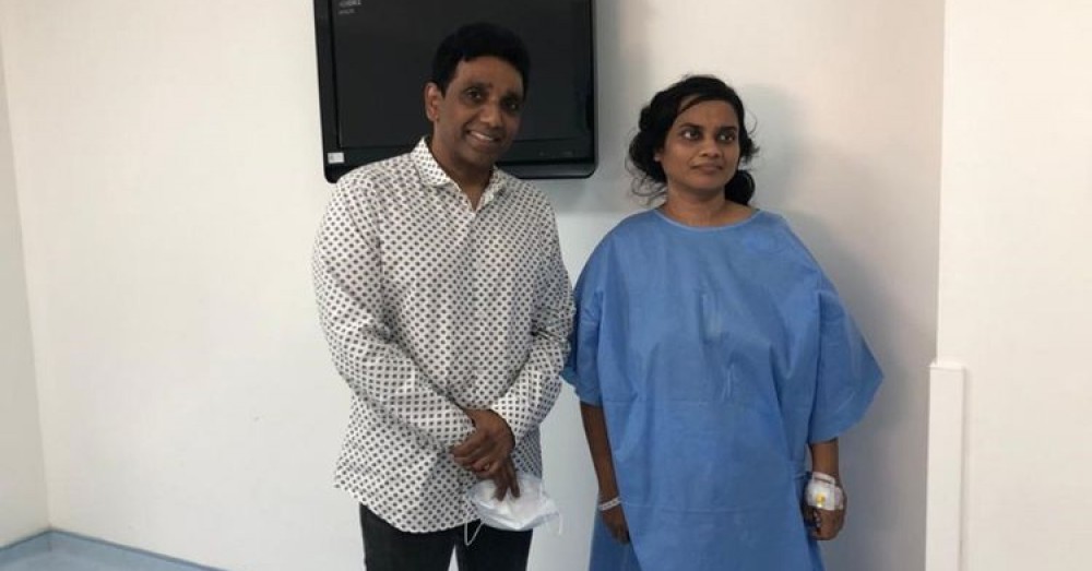
In the first-ever reported case of minimal invasive surgery to extract an 8cm cyst from the liver, a UAE surgeon was able to resect a portion of the liver of a 49-year-old woman, laparoscopically.
The patient, Christy Bai Gnanamai Parimala, a music teacher at a Fujairah-based school, had been suffering from abdominal swelling, breathlessness, pain and nausea and had lost her appetite for many days. The baseball-sized cyst contained live worms that were growing within the membrane after this music teacher had inhaled them from the faeces of her pet dog.
The three–hour keyhole surgery recently took place at the Thumbay Hospital in Fujairah.
What are Hydatid cysts?
Providing details of the case, Dr Raj Kumar, surgeon and specialist, minimal invasive surgery, told Gulf News: “Hydatid cysts are relatively rare in the UAE. These are caused when a person inhales or ingests microscopic larvae of the dog tapeworm (Echinococcus granulosus) excreted through the faeces of the animal. The larvae from a cyst that is attached to the liver continues to grow as the worms grow. This cyst can be fatal if it ruptures because as it releases the larvae into the blood stream. The liquid from the ruptured cyst can be highly toxic. Hydatid disease is fairly common in some parts of Saudi Arabia, Australia, parts of the United States and in the Indian subcontinent.”
The traditional procedure is to cut open the abdomen to extract the cyst. The minimally invasive surgery, carried out for cyst extraction in the Middle East, helps extract the cyst and parts of the infected liver through minor punctures on the abdominal area. This not only gives a better cosmetic outcome, but also accelerates healing, allowing the patient to resume a normal life within a week.
Cyst growing for 10 years
Parimala’s son, Jeremiah Caleb Henry said the cyst must have formed more than ten years ago. “My mother came here ten years ago to teach at the school. Prior to that, in Chennai, India, we had a pet German Shepherd dog and we presume that the source of this larvae was that pet which was eventually given away when she moved here.”
Henry said the cyst was spotted two-and-a-half years ago during a routine CT scan. “My mother had suffered from lower-back pain two-and-a-half years ago and during the scan, the doctor had spotted this tiny cyst, which he said was a common cyst that would go away with time. But for the last few months, my mother was complaining of something poking her stomach as the cyst was pressing from the liver on to her abdominal wall. Since she was in pain, discomfort, breathlessness and experienced loss of appetite, she underwent a CT scan — only to discover that the cyst had grown to be 8x7cm.”
After surgery, Parimala said that the pain had subsided and she could no longer feel the poking sensation in her tummy.
Share This Post


